
The AEC made the investment to obtain digital radiology in 2009 and have upgraded the system twice. The most recent upgrade was in 2021 with upgrade to IDEXX system which allows for improved sharing of images with your veterinarian. We have had ultrasound since 2005 and our new machine from 2020 is from Sonosite and is the same unit used in many university veterinary ER departments.
The main advantage of digital radiology is speed. We are able to obtain radiographic images very quickly without the need to process film. This advantage means that we can diagnose the problem faster to start treatment, or if no problems are found, then you and your pet get to go home sooner.
Digital imaging allows for multiple copies of the same radiograph or ultrasound image to easily be transferred to your veterinarian, a specialist for review or your personal computer if you had the interest in showing them to your family.
Many of our patients are very small. Digital radiology allows us to zoom in on specific areas and magnify their size by 600% without loosing detail quality. The digital program also lets us adjust the contrast, darkness and even reverse white and black. This decreases the need for retakes and allows a novel way to look at conventional radiology. “Hair-line” fractures and lung lesions can “jump-out” of the image to our trained veterinarians with this technology.
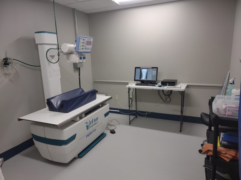
Our new radiology department is large and has an oxygen drop allowing the acquiring of diagnostics as an extension of our ICU.
Here are some examples of radiographs taken at the AEC. The detail is amazing here, and these images have been compressed significantly for website viewing.
Canine bloat is a common surgical problem. Digital radiology allows for rapid differentiation of a twisted stomach filled with air and a large dilated stomach filled with food. This is important because they are treated differently. The twisted stomach or “gastric dilation with volvulus” (GDV) is the disease that Marley died from in the movie “Marley and Me,” but with early detection and surgery most dogs will survive. Symptoms include gagging and producing white foam, restlessness, pain the belly and a very swollen abdomen. In the image below, the large black circle is the stomach. The white arrows point to where the stomach is actually twisted and the red arrow points to intestines dilated with air. The classic pattern below is often called the “double bubble,” “shelf sign,” “Popeye stomach,” or more professionally, “classic compartmentalization of the stomach associated with gastric dilatation and volvulus.”
 Canine “bloat” is a surgical emergency. |
Dogs eat the strangest things. Here are some examples of objects ingested by dogs which show up very well on radiographs. Many objects that dogs eat will either be vomited or pass in the stool, but often the foreign objects will get stuck as they go through the intestines and require surgery to have them removed. The most common symptom of a foreign body obstruction is vomiting, but they may also stop eating, become lethargic or act painful. Can you find the objects that were swallowed by these dogs?
 Intestinal obstruction from drawer pull |
 Intestinal foreign body with baby bottle nipple |
 A bottle cap in the stomach of a dog |
Trauma is a common cause of presentation to the ER. Some animals can severely injure themselves by simply jumping from the couch, others a deck or even a roof. When your pet is injured our digital radiology department allows us to quickly assess for fractures. One of these dogs was hit by a car and the other was stepped by a child in the house.
 Lateral view of metacarpal fractures |
 Ventro-dorsal view of metacarpal fractures |
 Radius and Ulna fracture of a dog. |
A perineal hernia is a fairly rare event. Surgery is typically required to repair it when it happens. In this case, the bladder has flipped backwards and squeezed out through the hernia. The bladder swelled as urine was produced and a bulge could be felt next to the tail base. After passing a catheter and draining the bladder it was able to be repositioned into the normal location. This provided the time to start treatment and have surgery performed.
 Bladder malpositioned with perineal hernia |
 Bladder replaced after perineal hernia |
Breathing problems are common in cats and dogs. There are many causes. The cat in the radiograph below was having difficulty breathing due to fluid build-up in the chest. This fluid build up was compressing the lungs. The lungs are the black triangle in the center of the picture. The rest of the chest is white due to the density of the fluid. The black triangle of lungs should fill the entire chest.
 Pleural effusion in a cat |